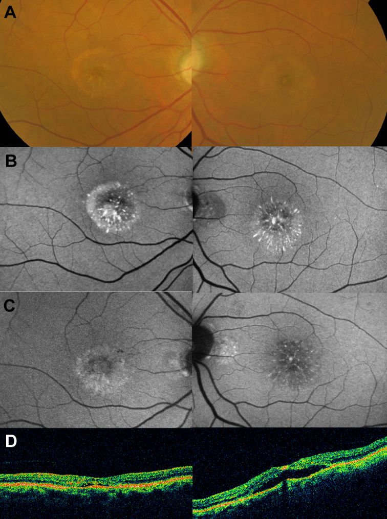
Figure 2.

Fundus appearance and autofluorescence findings 18 months following the initial visit (October 2006, see text). (A) Colour fundus photographs show disappearance of most of the subretinal material. (B) Autofluorescence imaging demonstrates small foci of very highly increased signal at the site of the macular lesions. In April 2008, a mildly increased autofluorescence signal was evident in the right eye at the macula surrounding a central area of mildly reduced atrial fibrillation; optical coherence tomography demonstrated a lack of foveal depression and a minimal amount of subretinal fluid (C). In the left eye, areas of reduced autofluorescence signal and few remaining foci of increased autofluorescence were seen; optical coherence tomography demonstrated a neurosensory retinal detachment (NSRD; D).
