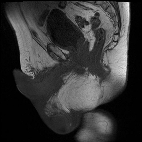
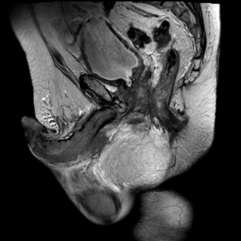
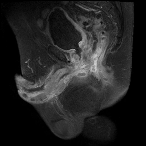
Figure 1.
Sagittal T1 (upper left), T2 (upper right) and postcontrast fat suppressed T1 (lower left) images through the midline of the pelvis demonstrate nodular metastasis and engorgement of the corpora cavernosa. Also note the irregularity of the prostate and thickening of the inferior bladder wall secondary to invasion by the primary adenocarcinoma of the prostate.


