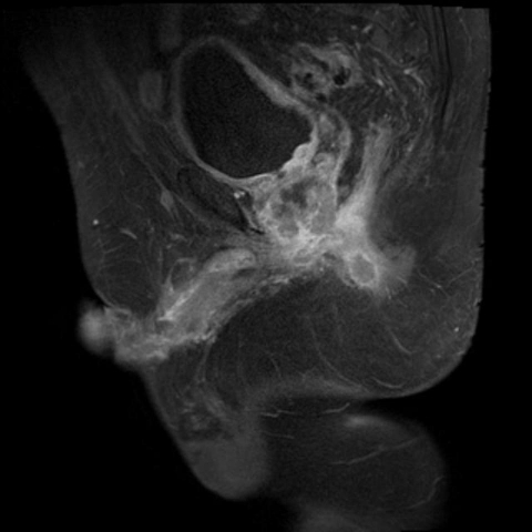
Figure 2.
Sagittal postcontrast fat suppressed T1 weighted image through the right lower pelvis demonstrates a peripherally enhancing nodule within the right ischiorectal fossa compatible with necrotic metastatic lymph nodes. Again seen is the irregularly enhancing primary prostatic mass with extension to the inferior bladder wall.

