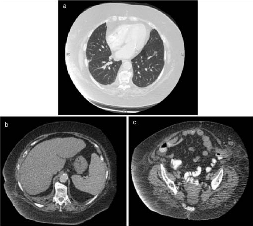
Figure 1.

Computerised tomography images of (A) the thorax at the level of the pulmonary trunk (lung window), showing both lungs free of metastases, (B) the abdomen at approximately the level of T10 showing the liver also free of metastases (note the mildly cirrhotic appearance of the liver, most likely the sequelae of non-alcoholic steatohepatitis) and (C) the pelvis at approximately the level of the mid sacrum showing no apparent enlargement of the uterus or lymphadenopathy.
