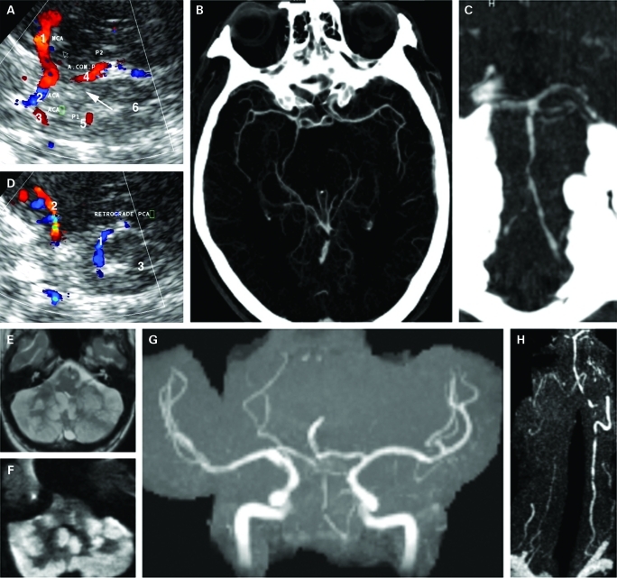
Figure 1.
A. On admission, transcranial colour-coded sonography, axial view of the mesencephalic image plane through the left temporal window: 1=retrograde filling of the left posterior cerebral artery (PCA) in blue away from the transducer (P1); 2=ipsilateral middle cerebral artery (MCA) with branches in blue. B. Transcranial colour-coded sonography, axial view of the mesencephalic image plane through the right temporal window: 1=right MCA (M1); 2=ipsilateral anterior cerebral artery (ACA) (A1); 3=contralateral ACA (A1); 4=filling of the right PCA (P2) via posterior communicating artery; 5=contralateral PCA (P1) now in red towards the transducer; 6=mesencephalic brainstem. Arrow marks missing right P1 segment due to either no flow, aplasia (fetal type) or hypoplasia. C. Maximum intensity projection of the circle of Willis from CT-angiography confirming fetal type origin of the right PCA and filling of the left PCA. D. Maximum intensity projection of the vertebrobasilar system showing multiple narrowing of the basilar and vertebral arteries. E. 7 day later, fluid attenuated inversion recovery (FLAIR) sequences showing massive ischaemia of the cerebellum. F. Diffusion-weighted MRI confirming acute cerebellar ischaemia. G. Time-of-flight MR-angiography (MRA) showing highly compromised flow in the vertebrobasilar system. H. Contrast-enhanced MRA showing multiple severe stenosis of the right vertebral artery, multiple distal high grade stenosis of the dominant left vertebral artery and basilar artery.

