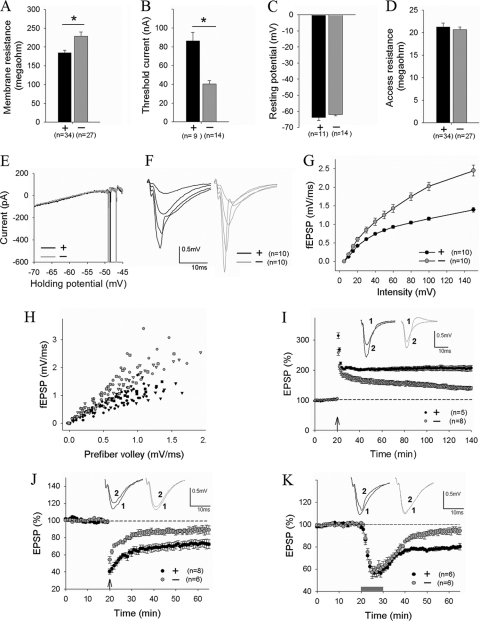
FIG. 7.
Enhanced basal synaptic strength and impaired synaptic plasticity in PAK1/PAK3 DK mice. (A to D) Whole-cell recordings of CA1 pyramidal neurons showing increased input resistance (A) and decreased current injections to elicit an action potential (B) but normal resting membrane potential (C), access resistance (D), and threshold potential to fire an action potential (E) in the DK neurons. (F to H) fEPSPs at CA1 synapse (F) and summary graphs (G and H) evoked by various stimulation intensities showing enhanced synaptic responses in the DK mice. (I, J, and K) fEPSP recordings showing significantly reduced NMDA receptor-dependent LTP induced by high-frequency stimulation (4 times at 100 Hz, lasting 1 s each, arrow) (I), diminished NMDA receptor-dependent LTD induced by 1-Hz stimulation (15 min) (J, arrow), and the abolishment of metabotropic glutamate receptor (mGluR)-dependent LTD induced by brief (10-min) application of 100 μM group I mGluR agonist DHPG (K, bar). n = number of slices or neurons.

