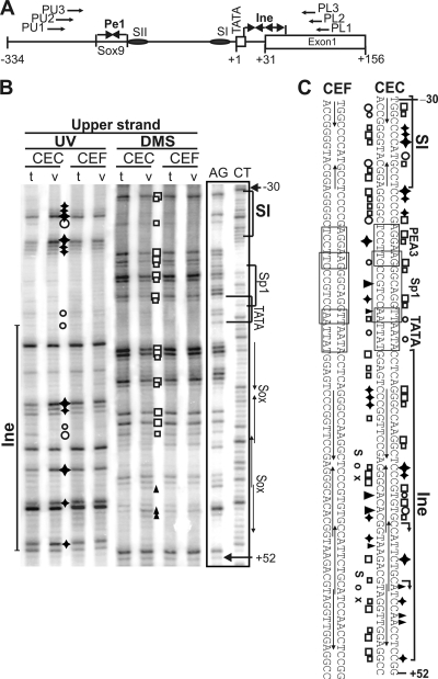
FIG. 4.
Tissue-specific occupancy of Ine1 and SI in genomic footprinting. (A) Schematic depicting the primers used in footprinting and the short promoter elements. (B) Footprints on the upper DNA strand. AG and CT are Maxam-Gilbert ladders. DNA from CEC and CEF cultures treated in vivo (v) with DMS (open boxes or solid triangles) or UV light (open circles or solid diamonds) is compared with the in vitro (t) DNA samples treated with these reagents after isolation from CEC and CEF. Differences in the modification patterns between in vivo and in vitro treatments appear as hyperactivities (solid diamonds or triangles) or protections (open circles or boxes), revealing specific in vivo DNA-protein contacts. (C) Summary of in vivo footprinting on both strands.

