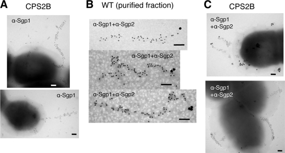
FIG. 4.
Immunogold staining of pilus structures of S. suis strains. (A) Immunogold-labeled Sgp1 (gold particle size, 10 nm) on the cell surfaces of strain CPS2B. (B and C) Double immunogold labeling with mouse anti-Sgp1 pAbs (α-Sgp1; gold particle size, 10 nm) and rabbit anti-Sgp2 pAbs (α-Sgp2; gold particle size, 20 nm) in the partially purified cell wall fraction of the wild-type (WT) strain (B) and on the cell surface of strain CPS2B (C). Scale bars = 0.1 μm.

