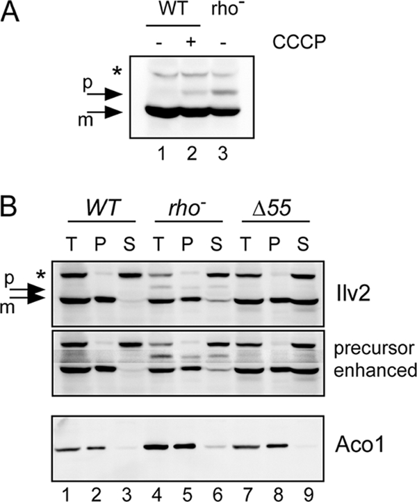
FIG. 2.

Accumulation of the Ilv2 precursor in the petite mutant. (A) Detection of Ilv2 by Western blotting. Lane 1, RKY2139 (WT); lane 2, RKY2139 (WT+CCCP, 40 μM, 4 h); lane 3, RKY2315 (rho−). (B) Fractionation of Ilv2. Cell extracts of the rho+ strain RKY2139 (lanes 1 to 3), the rho− strain RKY2315 (lanes 4 to 6), and the ILV2/ILV2-Δ55 strain RKY2491 (lanes 7 to 9) were fractionated by centrifugation. T, total fraction (lanes 1, 4, and 7); P, pellet fraction (lanes 2, 5, and 8); S, supernatant fraction (lanes 3, 6, and 9). Proteins were detected by Western blotting with anti-Ilv2 antibodies (upper two panels) and anti-aconitase antibodies (lower panel). In the second panel the region of the Ilv2 precursor was selectively enhanced. p, Ilv2 precursor; m, mature Ilv2. A background band is marked with an asterisk.
