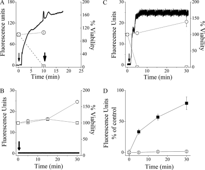
FIG. 4.
Aptitude of C16(ω7)K-β12 (4× the MIC) to disrupt the cytoplasmic membrane. Panels A to C show the simultaneous determination of bacterial viability and membrane depolarization using E. coli CI 16327 (A), E. coli ATCC 35218 (B), and S. aureus ATCC 29213 (C). Symbols in panels A to C: ○, untreated control; □, OAK-treated bacteria. Black traces show online monitoring of the fluorescence increase representing release of the potential-sensitive dye diSC3-5 in OAK-treated bacteria. Arrows point to the times of addition of the OAK. The second arrow in panel A points to the time for dermaseptin addition (4× the MIC). Panel D compares the PI uptake kinetics by OAK-treated E. coli ATCC 35218 (○) and S. aureus ATCC 29213 (▪).

