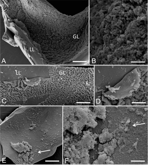
FIG. 3.
Ultrastructural effects of mefloquine on E. multilocularis metacestodes in vitro as visualized by SEM. Scanning electron microscopy (SEM) pictures were prepared for untreated metacestodes from in vitro-cultured parasites (A and B) and metacestodes treated for 2 h (C and D) and 6 h (E and F) with 24 μM mefloquine. Overviews of the vesicle tissue consisting of a laminated layer (LL) and a germinal layer (GL) are given in panels A, C, and E, whereas in panels B, D, and F, the focus is directed to GL cells detaching from the LL with increasing incubation times. Bars: panel A, 1,100 μm; panel B, 350 μm; panel C, 600 μm; panel D, 220 μm; panel E, 950 μm; panel F, 220 μm.

