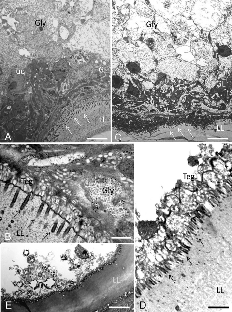
FIG. 4.
Ultrastructural effects of mefloquine on E. multilocularis metacestodes in vitro visualized by TEM. Transmission electron microscopy (TEM) pictures were prepared from in vitro-cultured parasites for untreated metacestodes (A and B) and metacestodes treated for 2 h (C), 12 h (D), and 48 h (E) with 24 μM mefloquine. Control vesicles showed a meshwork-like laminated layer (LL) with microtriches (indicated by arrows) protruding from the tegument (Teg) into the LL. The adjacent germinal layer (GL) consisted of densely packed cells (glycogen storage cells [Gly], undifferentiated cells [uc], and others). With increasing treatment time, glycogen storage cells were depleted (C) and the parasite tissue was no longer structurally intact (D); finally, the structural integrity of the GL broke down, microtriches were lost, and the LL acquired an amorphous appearance (E). Bars: panel A, 3.8 μm; panel B, 1.3 μm; panel C, 4.2 μm; panel D, 2.2 μm; panel E, 3.6 μm.

