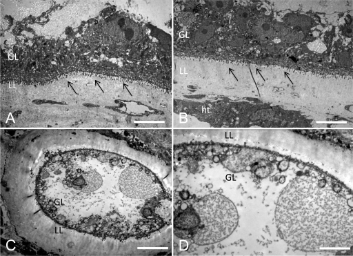
FIG. 6.
Ultrastructural effects of mefloquine on E. multilocularis metacestodes in vivo. Images from transmission electron microscopy (TEM) of metacestodes recovered from mice by the end of the in vivo treatment study are shown. (A) Untreated metacestodes show a well-defined laminated layer (LL) against host tissue (ht) and densely packed adjacent germinal layers (GL). (B) Metacestodes from animals treated orally with mefloquine present the same well defined structured and, at a higher magnification, undifferentiated cells and glycogen storage cells are visible, as well as well-defined microtriches (indicated by arrows). (C and D) The metacestodes recovered from mice treated intraperitoneally with mefloquine show a less-defined laminated layer as well as a thinner germinal layer. The undifferentiated cells are rounder in shape, and the glycogen storage cells are no longer visible. Bars: panel A, 4.2 μm; panel B, 4.2 μm; panel C, 5.4 μm; panel D, 2.6 μm.

