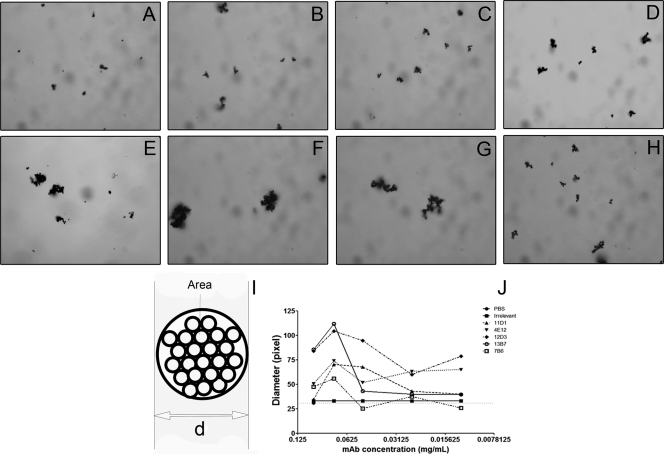
FIG. 2.
Agglutination of H. capsulatum cells measured by light microscopy and flow cytometry. (A to H) Microscopy of aggregates in which H. capsulatum is incubated with water (A), PBS (B), IgG isotype control (C), 11D1 (D), 4E12 (E), 12D3 (F), 13B7 (G), and 7B6 (H). MAb concentration, 0.075 mg/ml. (I) Depiction of the model used for ImageJ calculations of agglutination diameter (d). (J) Light microscopy shows that agglutination is dependent on concentration of MAbs used. Similar results were obtained by flow cytometry (data not shown). The results shown are representative of a minimum of three independent experiments.

