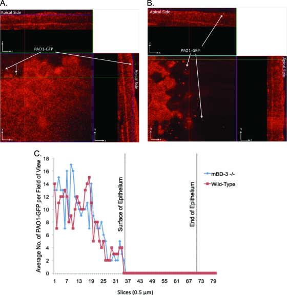
FIG. 7.
(A and B) Confocal reflection microscopy of mBD-3−/− (A) or wild-type (B) C57BL/6 mouse eyes at 7.5 h after inoculation ex vivo with ∼109 CFU P. aeruginosa strain PAO1-GFP (green). Eyeballs were carefully enucleated, rinsed with PBS, and then tissue paper blotted before bacterial challenge (see Materials and Methods). z-stack images were split into an orthogonal view to show x, y, and z planes of the intact corneal epithelium. After 7.5 h, bacteria showed equal levels of adherence to mBD-3−/− and wild-type corneas but still did not traverse the epithelium. (C) These data were confirmed by quantifying the bacterial distribution over the corneal epithelium (red indicates the reflection of corneal epithelial cells at 633 nm). Magnification, ×∼1,000.

