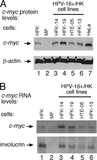
FIG. 3.
c-myc expression is altered in some HPV-16-immortalized integrated clonal human keratinocytes (HK). (A) c-myc protein levels were quantified by Western blot analysis of 30 μg of total protein extracted from clonal HPV+/HK cell lines (defined in Table 2) with integration at chromosome 10 (lane 3, clone HFK-14) and in chromosome 8 (c-myc) (lanes 4 to 6) compared to c-myc levels in primary HFK cells. Murine fibroblast (MF) cell extracts were used to demonstrate human c-myc antiserum specificity. (B) c-myc RNA levels quantified by Northern blot analysis of total RNA isolated from clonal HPV+/HFK cell lines and control cultures. The blot was stripped and rehybridized with a probe specific for cellular involucrin RNA. HeLa cells were used as a control for elevated c-myc levels compared to those of HFK (primary foreskin keratinocytes). Murine fibroblasts were used as a negative c-myc control.

