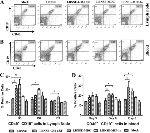
FIG. 5.
Recruitment and/or activation of B cells in the lymph nodes and blood after infection with rRABVs. BALB/c mice were infected i.m. with 1 × 10⁵ FFU of different rRABVs, and draining (inguinal) lymph nodes and blood were harvested after extensive perfusion at 3, 6, and 9 dpi. Single-cell suspensions were prepared, stained with antibodies against B cells and the B cell activation markers CD19 and CD40, and analyzed by flow cytometry. Representative flow cytometric plots of B cells are shown from the lymph nodes (A) and the blood (B). The detailed analyses for activated B cells (CD19+ and CD40+) at 3, 6, and 9 dpi are presented for lymph nodes (C) and blood (D). Asterisks denote significant differences between the indicated experimental groups (*, P < 0.05; **, P < 0.01).

