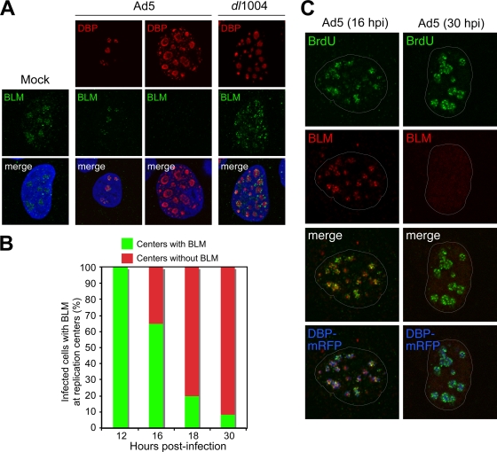
FIG. 3.
Accumulation of BLM at sites of viral replication. (A) HeLa cells were infected with wild-type Ad5 (MOI of 10) or E4 mutant dl1004 (MOI of 25). Localization of BLM was examined by immunofluorescence (14) after preextraction to remove soluble protein prior to fixation and compared to that for mock-infected cells (left). In uninfected cells, BLM localized to nucleoli and ND10 structures. In Ad5-infected cells, BLM was either located in discrete foci surrounding early viral replication centers (as detected with an antibody to the viral DBP) or undetectable in cells with large centers (representing late-stage infection). In cells infected with E4-deleted virus dl1004, all infected cells displayed BLM accumulated at viral centers. DAPI staining indicates the location of the nuclei in all merged images. (B) Quantification of infected cells with BLM at viral replication centers over a time course of wild-type Ad5 infection. At each time point, the relative number of cells with BLM in the two patterns demonstrated by the representative images shown in panel A was determined by examining 100 infected cells. (C) BLM at sites of active viral replication. HeLa cells were transfected with DBP-mRFP and, after 8 h, were infected with Ad5 (MOI of 10). At early (16 hpi) and late (30 hpi) stages of infection, cells were pulsed with BrdU for 30 min to label sites of DNA replication (46) and detected with a BrdU-specific antibody (Sigma). In preextracted cells, BLM could be detected adjacent to sites of ongoing viral replication. By 30 hpi, BLM had been degraded.

