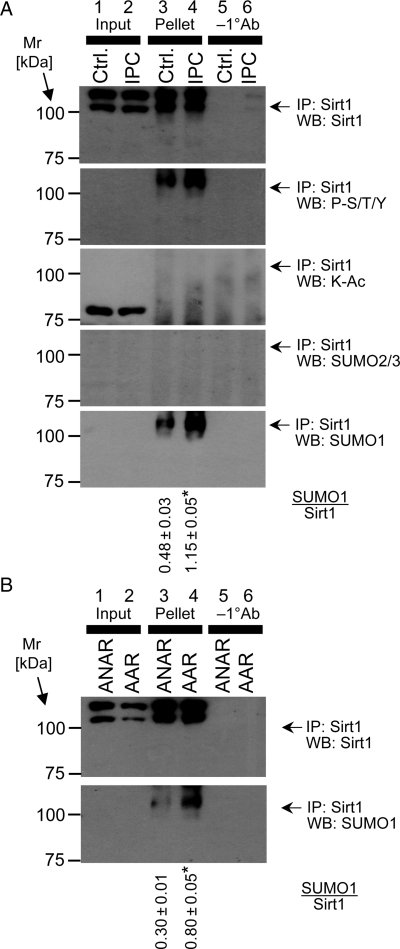
Figure 3.
PTMs of SIRT1 during IPC. (A) SIRT1 was immunoprecipitated from homogenates of perfused hearts subjected to control perfusion (Ctrl.) or IPC, without subsequent IR injury. In each blot, lanes 1 and 2 show the input to the immunoprecipitation (i.e. the tissue lysate), lanes 3 and 4 show the immunoprecipitated pellet, and lanes 5 and 6 show an experiment in which immunoprecipitation was performed without the primary (SIRT1) antibody. Panels (top to bottom) show western blots for SIRT1, phospho-Ser/Thr/Tyr, K-Ac, SUMO2/3, and SUMO1. (B) SIRT1 was immunoprecipitated from the AAR or ANAR of hearts subjected to IPC in vivo. Western blots for SIRT1 (upper panel) and SUMO1 (lower panel) are shown. Assignment of lanes 1–6 is as detailed for (A). In both panels, numbers to the left are molecular weight markers (kDa). Blots are representative of at least two independent immunoprecipitation experiments in the in vitro or in vivo condition. The ratio of SUMO1/Sirt1 was calculated and is shown below the blots (mean ± standard deviation). *P < 0.05 (Student's t-test) between control and IPC. Loading controls (representative Ponceau S stained membranes) for all blots in are shown in Supplementary material online, Figure S6.

