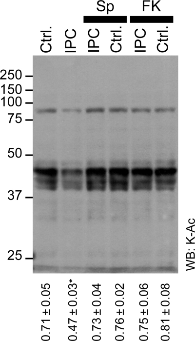
Figure 4.
Effect of SIRT1 and Nampt inhibition on lysine acetylation in IPC. Cytosolic fractions from hearts subjected to control perfusion (Ctrl.) or IPC, in the absence or presence of splitomicin (Sp, 10 µM) or FK-866 (FK, 1 µM) were western blotted for K-Ac. Representative blot is shown, numbers to the left are molecular weight markers (kDa). Densitometry was performed in the range 25–100 kDa, normalized to protein across the same molecular weight range (representative Ponceau S stained membrane is shown in Supplementary material online, Figure S7). The ratio of K-Ac/Protein was calculated and is shown below the blots (mean ± SEM, n ≥ 5). *P < 0.05 (ANOVA) between the indicated group and all other groups.

