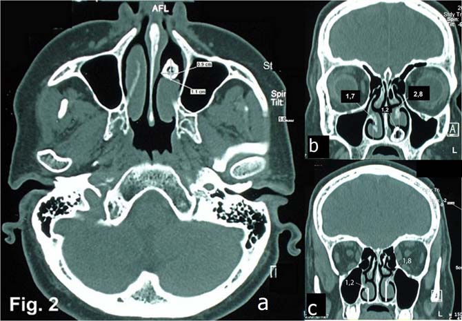
Figure 2.

Axial CT image (A) shows a radiopaque object with a radiolucent core (anteroposterior, sagittal dimensions were 9×11 mm) in the anterior third of the left nasal cavity. Atrophy of mucosa and os turbinalis is visible. Coronal CT views of the anterior (B) and middle (C) third of the inferior nasal meatus show the height difference between the left (2.8 cm/1.8 cm) and right (1.7 cm/1.2 cm) meatus. No other anatomical alterations were detected.
