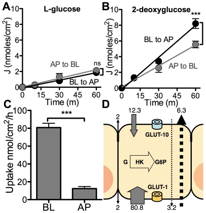
Figure 3. Airway epithelia can deplete airway surface liquid glucose.
(A and B) Basolateral to apical (BL to AP) and apical to basolateral (AP to BL) fluxes of L-[1-14C]glucose (A) or 2-deoxy-d-[1-14C]glucose (B) were measured in cultures of human airway epithelia over 1 hour. Data shown as mean ± s.e.m. n = 6 samples per group. (***: p<0.0001, ns: p ≥ 0.05). (C) Basolateral (BL) and apical (AP) uptake of 2-deoxy-d-[1-14C]glucose (2-DOG) in cultures of human airway epithelia were measured over 1 hour. Data shown as mean ± s.e.m. n = 6 samples per group. (***: p<0.0001). (D) Data from Figures 3 A, B and C are integrated into a model of glucose transport in human airway epithelia. Data in nmol/h. Black solid arrow represents paracellular diffusion of glucose. Gray solid arrows represent 2-DOG uptake capacity across the apical (top) and basolateral (bottom) membranes. Dotted arrows represent transcellular bidirectional fluxes of 2-DOG (adjusted by subtracting corresponding L-glucose fluxes). GLUT-10 is present in the apical membrane and GLUT-1 is present in the basolateral membrane. Intracellular glucose (G) is phosphorylated by hexokinase (HK) for subsequent glycolysis. G6P = glucose-6-phosphate.

