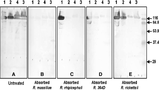
Figure 1.

Western blotting assay and cross-absorption studies on serum from dog B, California. Western blotting was performed with serum collected on 4/20/2009 and 15 μg of rickettsial protein per lane (A). Treatment of serum with antigen diluents (A), and Rickettsia massiliae (B), R. rhipicephali (C), 364D Rickettsia (D) and R. rickettsii (E) antigens, followed by Western blotting on the resulting treated serum samples, was performed according to the procedure described in the Materials and Methods. Antigens were loaded on the gel in the following order: lane1, R. massiliae AZT-80; lane 2, R. rhipicephali CA871; lane 3, 364D Rickettsia; lane 4, R. rickettsii Bitterroot. Positions of prestained broad range sodium dodecyl sulfate-polyacrylamide gel electrophoresis standards (Bio-Rad, Hercules, CA) are indicated on the sides of the blots. All serum samples were tested by Western blotting at a final dilution of 1:5,000.
