Figure 3.
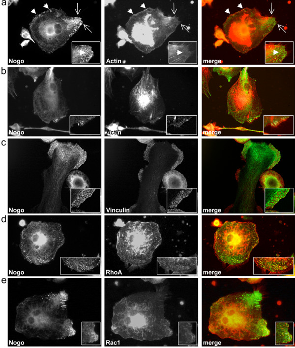
Nogo-B and cytoskeletal structures in monocyte-derived macrophages. (a) Partial co-localizations of Nogo-B and actin in punctate and elongate peripheral cell structures (arrowheads) and at the base of filopodia (arrows) with enlarged detail in inlay. Note that Nogo-B is not always present at the base of filopodia. (b) Nogo-B expression at the base of filopodia (see inlay). Actin stress fibers are not positive for Nogo-B. (c) Nogo-B is excluded from focal and podosomal adhesion sites (see inlay) (d) Intracellular, longish staining patterns of Nogo-B partially co-localize with RhoA (see inlay) (e) Nogo-B is present in Rac1 positive peripheral membrane ruffles (see inlay). Cells shown are representative of regularly observed staining patterns/co-localizations in independent macrophage cultures of six different donors. All images were acquired by conventional fluorescence microscopy using a DMI 4000B microscope and Application Suite V3.1 by Leica and represent cells cultured in 24-well cell culture plates.

