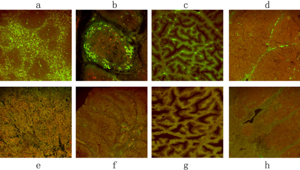
Figure 3.

The immunofluorescence distribution of DPV gE antigen on immunological organs. The tissue sections were stained with indirect immunofluorescent assay at 24 h post-infection. The positive signal of the gE antigen appeared yellow-green, and the positive staining was widely distributed in a to d (spleen, BF, Harderian gland, thymus); and no positive signals were detected on the spleen, BF, Harderian gland, thymus of mock-infected ducks (e to h).
