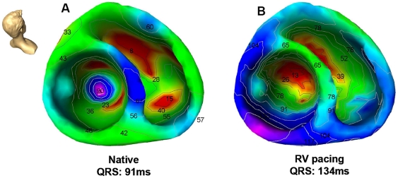
Figure 2. Color-coded electroanatomic activation map of the right and left ventricle in a control patient (no structural heart disease) during A) intrinsic sinus rhythm (native) and B) right ventricular (RV) pacing.
Red color illustrates earliest activation, blue color illustrates area of late ventricular activation. Head icon indicates point of view.

