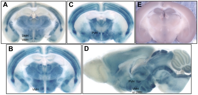
Figure 5. Distribution of Rcan2 in the hypothalamus.
(A-D) X-gal staining of brain coronal sections (A–C) and sagittal sections (D) revealed that Rcan2 was highly expressed in the dorsomedial (DMH), ventromedial (VMH) and paraventricular (PVH) hypothalamic nuclei. (E) X-gal staining of wild-type controls.

