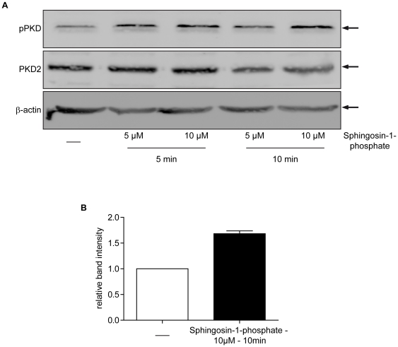
Figure 2. PKD2 is activated by sphingosine-1-phosphate (S1P) in C2C12 cells.
Subconfluent C2C12 cells were stimulated as indicated in the figure with (S1P) and further processed for immunoblot analysis for PKD2 and phospho-PKD (Ser 744/748). (A) One representative western blot for PKD2 and phospho-PKD (Ser 744/748) out of three independent experiments is shown. β-actin served as loading control. (B) Quantification of band intensity in the shown immunoblot of phospho-PKD expression relative to PKD2 in S1P-stimulated C2C12 cells. Values include 10 min values of 10 µM S1P treatment from all three experiments.

