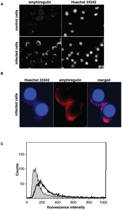
Figure 3. N. gonorrhoeae infection causes an increase of membrane bound amphiregulin that co-localizes with the site of bacterial adherence.
Non-permeabilized Me-180 cells were infected with N. gonorrhoeae for 6 hours. Amphiregulin was detected by using polyclonal antibodies against amphiregulin (MBS220633), followed by secondary IgG antibodies conjugated with fluorofor Alexa 488 nm. (A) Representative immunofluorescence images visualizing the protein levels of amphiregulin on the plasma membrane in control cells and infected cells. Cellular DNA and bacterial DNA was stained with the cell-permeable dye Hoechst 33342. (B) High magnification images of infected Me-180 cells. Image showing the co-localization between surface bound amphiregulin (red) and bacterial DNA staining (blue). (C) Flow cytometry histogram showing the protein levels of plasma membrane bound amphiregulin in non-permeabilized cells. Amphiregulin staining on uninfected control cells is shown in grey while staining of infected cells is shown with a black line. The histogram is representative of two independent experiments.

