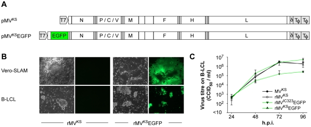
Figure 1. Generation and growth of rMVKSEGFP.
(A) Plasmids generated after RT-PCR, cloning and sequencing of MV RNA isolated from MVKS-infected PBMC. pMVKS is a full-length plasmid containing the complete antigenome of MVKS and pMVKSEGFP was modified by the insertion of an ATU at the promoter proximal position containing the ORF encoding EGFP. (B) rMVKS and rMVKSEGFP were rescued from Vero-SLAM cells and passaged in B-LCL. Fluorescence microscopy confirmed high levels of EGFP expression in rMVKSEGFP infected cells. (C) Growth curves of MVKS, rMVKS, rMVKSEGFP and rMVIC323EGFP in human B-LCL. Virus was harvested 24, 48, 72 and 96 hours post infection, CCID50 was determined in an endpoint titration test. Measurements shown are averages of triplicates ± SD. Key: h.p.i.: hours post infection.

