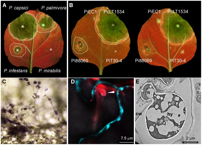
Figure 1. N. benthamiana shows varying degrees of susceptibility to Phytophthora species and P. infestans isolates.
Detached leaves of N. benthamiana were infected with spore solution droplets (marked as X) of Phytophthora capsici LT1534, P. palmivora 16830, P. infestans 88069 and P. mirabilis PIC99114. (A) or P. infestans isolates (B) and using P. capsici as a reference. Photographs were taken 3 days post inoculation under UV illumination. Lines mark infected areas, dotted lines mark the border between necrotic tissue and biotrophic tissue. P. infestans is able to produce sporangia on N. benthamiana 8 days post infection (C). P. infestans isolate 88069td (expressing tdTomato red fluorescent protein) formed digit like haustoria (arrowhead) that invaginated the N. benthamiana cell membrane labelled by transient Agrobacterium tumefaciens mediated expression of a membrane localised cyan fluorescent protein at 3 dpi (D). Haustoria were also observed by electron microscopy (E). h, haustorium; cw, cell wall; hy, hypha.

