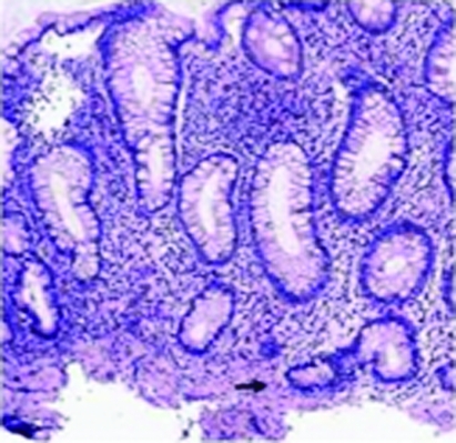
Figure 1.
Colonoscopy examination showed multiple linear ulceration with “cobble stoning” appearance starting from descending colon to caecum sparing the rectum. Multiple biopsies were taken histologically showing sections of colonic mucosa with crypt branching and cryptitis in caecum, ascending, transverse colon. There was no cryptitis in descending, sigmoid colon and rectum with some crypt branching and no granuloma in all sections.

