Fig. 3.
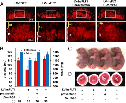
Pravastatin and mPGF restored impaired placentation and IUGR in the preeclampsia. (A–D) Placenta-specific hsFLT1 expression impaired placental bed development and caused IUGR. (A) Impaired vasculogenesis in the placenta was restored by pravastatin treatment and mPgf expression. Placentas were collected at E13.5, and vascular endothelial cells were stained with anti-CD31 antibody so that the region with more blood vessels appears in red. The labyrinthine area indicated with white box in Upper is magnified in Lower. la, labyrinthine layer; sp, spongiotrophoblast layer. (B–D) Fetuses and placentas collected by Caesarian section at E18.5. (B) Average fetal and placental weight. There are significant differences among the values labeled with different lowercase letters in the same color (P < 0.05). (C) Live and healthy pups were obtained in all groups. Malformation in gross appearance was not observed after pravastatin treatment. Because the difference in weight is ≈20% and is ≈6% in each dimension, it is difficult to see the weight differences in the photographs. (D) Placentas. Photos were taken from the fetal side.

