Fig. 1.
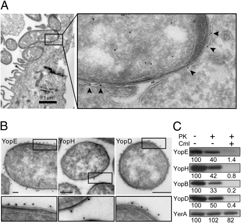
YopE is evenly distributed in the bacterium-target cell interface during infection of HeLa cells and Yops are found on the surface of Y. pseudotuberculosis before target cell contact. (A) Immunoelectron micrograph showing localization of YopE in Y. pseudotuberculosis during infection of a HeLa cell. Arrowheads indicate YopE detected on the surface of or within the plasma membrane of the target cell. (B) Immunoelectron micrographs showing surface localization of YopE, YopH, and YopD on Y. pseudotuberculosis before target-cell contact. (Scale bars, 100 nm for YopE and YopH; 200 nm for YopD.) (C) Western blot analysis of YopE, YopH, YopB, YopD, and the cytoplasmic T3SS chaperone YerA (SycE) in Y. pseudotuberculosis treated with chloramphenicol (Cml) or proteinase K (PK). Relative signal intensities are indicated below each panel.

