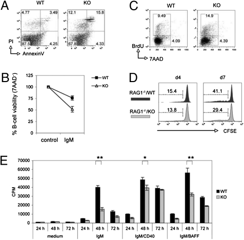
Fig. 2.
B-cell apoptosis and proliferation. (A) Increased apoptosis of resting splenic B cells. FACS of PI versus Annexin V staining of B220+-gated splenocytes. (B) Increased BCR-mediated apoptosis. Purified splenic B cells were left untreated (medium) or stimulated for 24 h with 5 μg/mL anti-IgM (IgM). Viability was determined by 7AAD staining and FACS. (C) Increased BrdU incorporation in vivo. WT and KO mice drank water containing 1 mg/mL BrdU for 3 d and B220+ splenocytes were analyzed by intracellular staining and FACS (BrdU vs. 7AAD). (D) Inhibition of homeostatic expansion. Splenic B cells (5 × 10⁶) were labeled with 5 μM CFSE and injected into 6-Gy–irradiated RAG1−/− mice. At 4 and 7 d after injection, CD19+-gated splenocytes were analyzed by FACS. Data are the percentage of B cells showing CFSE decay relative to total B cells. (E) Reduced [3H]thymidine incorporation. Splenic B cells (1 × 10⁵) were left untreated (medium) or stimulated with 5 μg/mL anti-IgM (IgM), 5 μg/mL anti-IgM plus 5 μg/mL anti-CD40 (IgM/CD40), or 5 μg/mL anti-IgM plus 5 ng/mL BAFF (IgM/BAFF). [3H]thymidine incorporation was assessed at 24, 48, or 72 h after seeding after a 9 h pulse (1 μCi). *P < 0.05; **P < 0.005.

