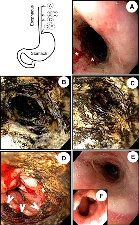
Fig. 1.
Black esophagus: Images of the esophagus acquired during upper endoscopy show linear ulceration (asterisk) of the mucosa lining the proximal half of the esophagus (a), and circumferential greenish-black appearance in the distal half of the esophagus (b, c). This segment of black discoloration ended abruptly 1–2 cm proximal to the squamo-columnar junction (d, arrowheads); appearance consistent with a diagnosis of acute necrotizing esophagitis (ANE). Image acquired during a follow-up endoscopy shows complete replacement of the black segment with normal mucosa within 6 weeks (e), and development of distal esophageal strictures within 4 months (f). (Color figure online)

