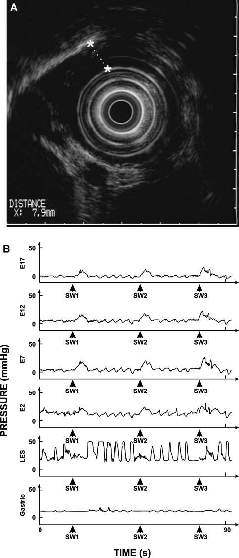
Fig. 2.
Achalasia Cardia. a Endoscopic ultrasound showed wall thickening of the thoracic esophagus, primarily involving the muscularis propia with a maximal thickness of ~8 mm (stars with interrupted line). b Esophageal manometry record shows three swallow-induced events (SW1, SW2, and SW3) in the esophagus, LES (lower esophageal sphincter), and stomach. Note that each swallow is associated with a simultaneous pressure wave in the esophagus and there is no LES relaxation, findings consistent with a diagnosis of achalasia esophagus

