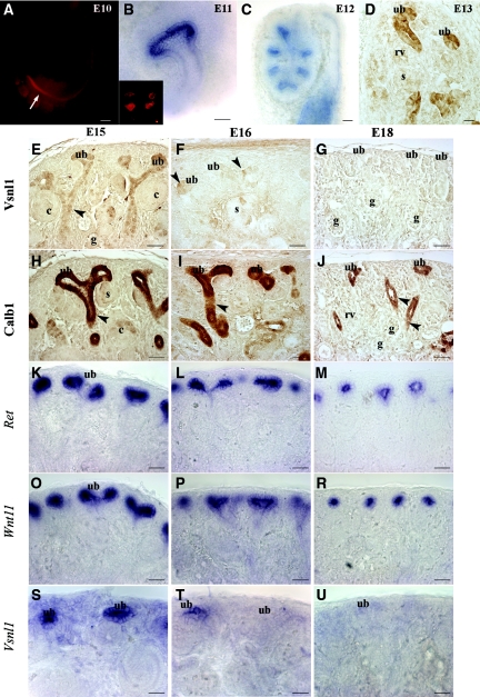
Figure 2.
Vsnl1 is required for early ureteric branching and is not required for the late stage of kidney development. Whole-mount antibody staining of Vsnl1 at E10 shows high levels at the caudal segment of WD close to cloaca (arrow in A). In situ hybridization (B) of E10.5 kidneys and protein labeling (B, inset) show Vsnl1 expression in the tips of the UB. (C) Vsnl1 mRNA in E12 kidneys is tip specific. (D) At E13, Vsnl1 protein levels are high in the UB tips and terminal branches but absent from renal vesicles (rv) and S-shaped bodies (s). (E) At E15, Vsnl1 protein is highly expressed in the tips of the UBs, and gradually lowering expression is seen throughout the terminal stalk region (arrowhead). No expression is observed in comma shaped bodies (c) or glomeruli (g). (S) In contrast to the protein, the Vsnl1 mRNA remain specifically expressed in the UB tips at E15. (F and T) At E16, Vsnl1 starts to decline, and few UB cells are still expressing at low levels the protein (F, arrowheads) and mRNA (T). (G and U) By E18 Vsnl1 protein and transcript expression are absent from the UB tips. (H through R) For comparison, the expression of Calb1 (H through J), Ret (K through M), and Wnt11 (O through R) is shown at E15, E16, and E18. Scale bars, 50 μm.

