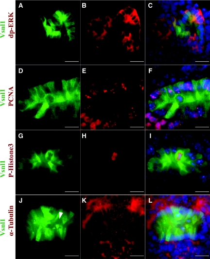
Figure 4.
The mosaic expression pattern of Vsnl1 protein does not correlate with cell cycle or ERK activity. (A through C) Double immunolabeling of E12 kidney sections with anti-dp-ERK1/2, a proliferation marker, and vsnl1 shows no correlation between the two molecules. (D through F) To visualize the synthesis phase of the cell cycle, double labeling with antibodies for Vsnl1 (D and F) and proliferating cell nuclear antigen (E and F) was performed. (G through L) The mitotic cells were visualized by double immunostaining with antibodies against Vsnl1 (G, I, J, and L) and P-His3 (H and I). Vsnl1 expression is high in P-His3-positive cells but also in some P-His3-negative cells. α-Tubulin marks the mitotic spindles (K and L). The arrowhead in J marks a cell in mitosis showing low levels of Vsnl1. DAPI (blue) marks the nuclei in C, F, I, and L). Scale bars, 10 μm.

