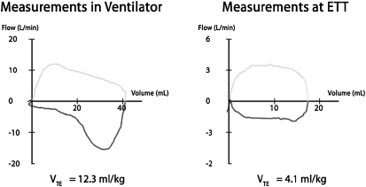
Figure 5.
Measurement from a 4.0-kg infant with a cuffed endotracheal tube in pressure-control mode with tube compensation active. On the left, flow-volume measurements are made at the ventilator with compensation for tubing compliance. There is “overshoot” of flow measurements causing volumes to be larger and giving the expiratory portion of the flow-volume curve (below the horizontal axis) a pattern of obstructive airways disease. Vt is 12.3 ml/kg. On the right, measurements are made at the endotracheal tube connector within a minute of the left panel. Here, the flows and volumes are much lower with Vt now one-third at 4.1 ml/kg. The flow pattern on the expiratory limb now resembles that of normal airways. ETT = endotracheal tube; VTE = exhaled tidal volume.

