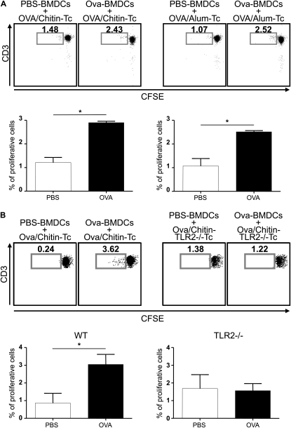
Figure 5.
Effects of chitin in ovalbumin (OVA)-induced T-cell proliferation ex vivo. (A) Cocultures were prepared containing T cells (Tc) from wild-type (WT) mice immunized with OVA complexed to chitin (left pair) or alum (right pair) and OVA- or vehicle control–stimulated bone marrow–derived dendritic cells (BMDCs) from WT mice. (B) Identical cocultures were prepared using Tc from Toll-like receptor 2 (TLR-2)-sufficient and -deficient (TLR2−/−Tc) mice. Proliferation of carboxyfluorescein diacetate succinimidyl ester (CFSE)-labeled CD4+ T cells (vs. CD3 expression) was analyzed using flow cytometry at Day 5. Daughter cell generation is shown in the square box compared with undivided parental generation and is also represented as a percent of proliferative cells (histograms). One representative out of five independent experiments performed is shown. Results are presented as the mean ± SEM of a minimum of six mice per group. *P < 0.05.

