Fig. 1.
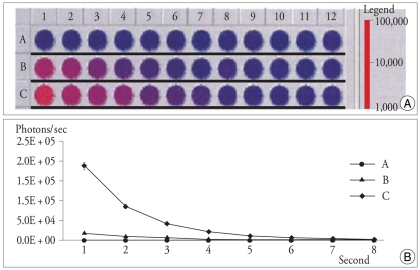
Two-fold serial dilutions were used to determine the luciferase activity by luminometry : Luminometer image of 96 well plate showing luciferase expression tests on progressive dilutions from 105 (row B), 106 (row C) of BMSC, and 106 (row A) of 293 T cells as a control. Each wells are containing 150 µL of PBS and 150 µg/g of luciferin was added followed by incubation period of 15 min at room temperature and measured with a second/well integration time (A). Plotting of luciferase activity examined by luminometer discloses two-fold decrease of photons counts per second in different initial cell numbers of BMSC (B : 105 cells, C : 106 cells). 293 T cells (A) show no detectable luminometric photon signal (B).

