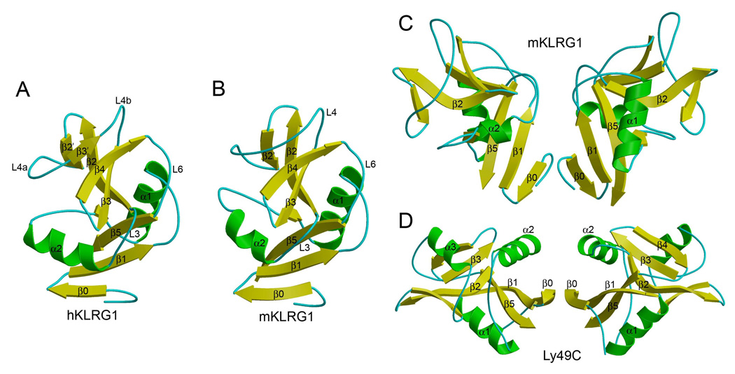
Figure 2. Structure of KLRG1.
(A) Ribbon diagram of human KLRG1, as observed in the hKLRG1–hEC1 complex. Secondary structure elements are labeled. α-helices are colored in green, β-stands in yellow, and loops in cyan.
(B) Structure of mouse KLRG1 in unbound form.
(C) Mouse KLRG1 homodimer, as observed in the mKLRG1–hEC1 complex. This dimer was not observed in structures of mKLRG1 alone or in the hKLRG1–hEC1 complex.
(D) Structure of the Ly49C homodimer (Protein Data Bank accession code 3C8J). Secondary structure elements are colored as in (A).

