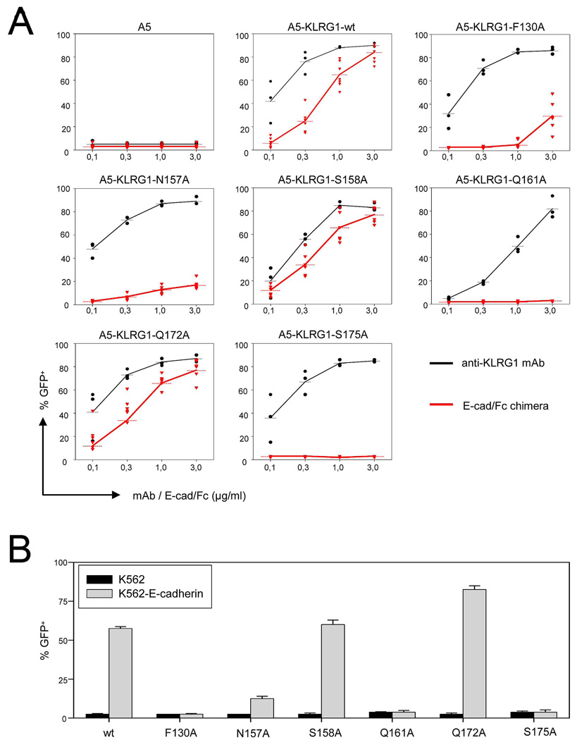
Figure 7. Mutational Analysis of the KLRG1–E-Cadherin Interface.
(A) A5-KLRG1 reporter cells carrying the indicated alanine substitutions in the extracellular domain of human KLRG1 were cultured in plates pre-coated with different concentrations of anti-KLRG1 mAb (black lines) or E-cadherin-Fc chimera (red lines). Cells were harvested after 17 h and induction of GFP expression was determined by flow cytometry. Dots represent values from individual assays and data from at least three independent experiments are shown.
(B) A5-KLRG1 reporter cells were co-cultured with parental (black bars) or E-cadherin-transduced (gray bars) K562 cells. GFP expression was determined after 17 h. Results are shown as mean values including SEM derived from three independent experiments.

