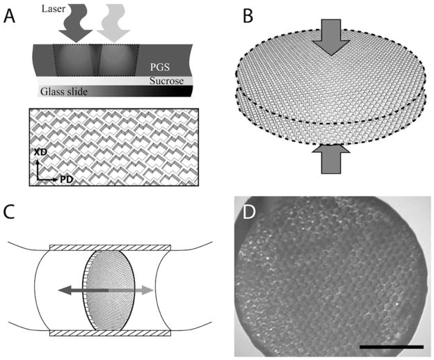
Figure 1.
Method. (A-C) PGS membranes were (A) laser microablated to make one-layered scaffolds with accordion-like honeycomb pores, (B) stacked and laminated to produce two-layered scaffolds, and (C) seeded with heart cells and cultured with bi-directional interstitial perfusion. (D) Representative phase contrast micrograph of a 7-day construct, scale bar: 2 mm.

