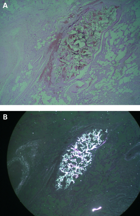
Figure 2.
A. Photomicrograph of a cross-section of the embolised arteriovenous malformation in the cheek showing extensive microthromboses (original magnification ×40). B. Photomicrograph of the same cross-section of the malformation visualised under polarised light showing the same foreign body particles among thrombi (original magnification ×40).

