
Figure 1.
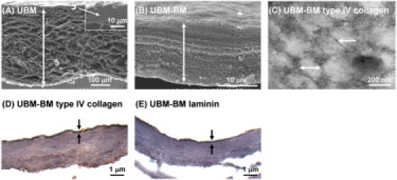
Characterization of UBM-BM and UBM. As model BM and stromal matrices, we characterized two ex vivo tissue scaffolds: UBM-BM and UBM. (A) Scanning EM image (×180) of a UBM cross-section reveals a thick stromal layer of connective tissue (double-headed arrow) underlying a thin layer that includes the BM (bracket and inset). (B) UBM-BM was created by mechanically delaminating porcine bladders to a further extent than UBM to obtain a very thin layer of connective tissue (double-headed arrow) underlying the BM (∗) that was equivalent to the top portion of UBM (bracket and inset of A) as shown in a cross-sectional scanning EM image (×8000). (C) After salt extraction of noncollagenous components, transmission EM imaging (×110,000) revealed that UBM-BM contains filaments (single-headed arrow) and 100–200 nm diameter pores (double-headed arrow) consistent with the polygonal type IV collagen network of a BM. The integrity of the BM of UBM-BM was confirmed histologically with continuous positive staining for (D) type IV collagen and (E) laminin (between the arrows).