
Figure 7.
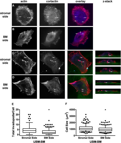
Invadopodia formation is enhanced on the stromal side of UBM-BM. To determine whether the stroma or BM is more permissive for formation of invadopodia, CA1d breast cancer cells were cultured overnight on the stromal or BM side of UBM-BM. Invadopodia (arrows) were identified by colocalization of actin (red) and cortactin (blue) for quantification on the (A) stroma or (B) BM using wide-field fluorescence imaging and confirmed with confocal imaging (C and D, z-stacks). The matrix surfaces were identified by collagen autofluorescence (green). (E) Quantitation of invadopodia formation on the stromal and BM sides of UBM-BM reveals a statistically significant increase in the total invadopodia/cell in cells cultured on the stroma. (F) Cell size is also statistically greater on the stroma. Data are presented as box and whisker plots with solid lines indicating medians, whiskers representing 95% confidence intervals, and dots representing outliers. ∗p < 0.05 for BM and stromal side comparisons; n ∼ 300 cells from two independent experiments.