
Fig. 2.
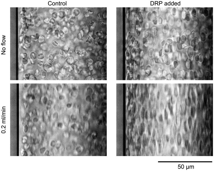
RBC suspension in a 100-μm wide rectangular straight channel. Addition of 10 ppm PEO-4500 caused a perceptible alignment of RBC in the non-flowing samples, and a dramatic elongation of RBC aligned with the flow direction (top to bottom) at 0.2 ml/min, Re ∼ 20. The PEO effectively eliminated the cell-depleted layer observed in the control sample. The slight difference between Control and DRP cell alignment at the no flow condition was due to the fact that the pictures were taken right after flow was stopped before the RBC started to sediment.