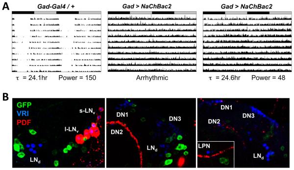
Figure 6. Hyper-exciting GABAergic neurons causes behavioral arrhythmicity.
(A) Representative actograms of heterozygous control Gad-Gal4 / + fly (left) and two flies with Gad-Gal4 expressing UAS-NaChBac2 (Gad > NaChBac2, center and right). ClockLab marked the Gad > NaChBac2 center fly as arrhythmic while the fly on the right as having a period of 24.6hr and a low power rhythm. Rhythmic Gad > NaChBac2 flies had weaker power rhythms then control flies (p<.001).
(B) Localization of Gad-Gal4 expression using a UAS-CD8::GFP reporter in three individual brains stained at ZT20 with antibodies to GFP (green), VRI (blue, marks all clock neurons) and PDF (red). Although some GFP+ neurons are close to VRI+ clock neurons, there is no obvious co-localization of VRI and GFP. Images are representative of 13 brain lobes.

